
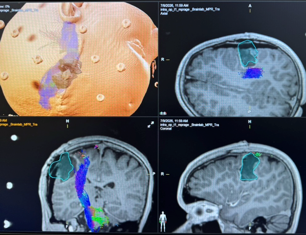
Surgery for brain tumors in children places the highest demands on precision and safety. At the Department of Neurosurgery at the Medical University of Vienna and Vienna General Hospital (AKH Vienna), advanced techniques such as computer-assisted navigation, robotics, augmented reality, and intraoperative imaging are used. These technologies are intended to help remove tumors as completely as possible while preserving important neurological functions.
The developing brain of a child is still undergoing significant changes. Procedures involving this sensitive organ therefore require particularly careful planning and execution. In addition, many brain tumors are located in close proximity to areas responsible for movement, speech, vision, or other essential functions. Others are situated deep within the brain or are difficult to distinguish from healthy tissue due to their characteristics.
For many pediatric brain tumors, surgery is a crucial component of treatment. Its goals are to safely remove as much tumor tissue as possible, obtain a reliable diagnosis, and at the same time protect healthy brain tissue, nerves, and blood vessels. Modern technological techniques support neurosurgeons in achieving this balance with increasing precision.
Navigation as an Orientation System in the Operating Room
Neuronavigation works similarly to a highly precise navigation system. Before surgery, magnetic resonance imaging and, when necessary, computed tomography scans of the child are converted into a three-dimensional model. During the operation, the exact position of the instruments can be continuously correlated with these imaging data.
This makes it possible to plan the most favorable and least invasive route to the tumor while deliberately avoiding important structures. Navigation provides particularly valuable orientation when dealing with small, deeply located, or anatomically difficult-to-reach tumors. However, it does not replace the experience of the surgical team; rather, it complements that expertise with additional spatial information.
Robotics for Particularly Precise Procedures
Robotic systems are used primarily when instruments need to be guided with a very high degree of accuracy along previously planned trajectories. This includes, for example, stereotactic tissue sampling from deeply located tumors or the precise placement of probes and catheters.
The robot does not perform the surgery autonomously. Planning, control, and supervision remain entirely the responsibility of the neurosurgeons. The robot serves as a highly precise assistive system that can reliably implement planned access routes. This allows certain procedures to be performed through very small openings, thereby reducing the physical burden on the child.
Augmented Reality Makes Hidden Structures Visible
Augmented reality combines the real surgical field with digital imaging information. Three-dimensional representations of the tumor, important blood vessels, or functionally significant areas of the brain can be superimposed within the surgical team’s field of view.
In this way, augmented reality can provide something resembling a view beneath the visible surface of the brain. The spatial relationships between the tumor and healthy structures can be understood more easily. This can be particularly helpful with complex tumors, supporting orientation and helping the surgical team identify critical areas at an early stage.
Some of these applications are still being further developed. However, the combination of navigation, surgical microscopy, robotics, and augmented reality is considered a promising approach toward increasingly individualized surgical procedures.
Monitoring During Surgery
One particular challenge is that the brain can shift slightly during a surgical procedure. Images acquired before surgery may therefore no longer correspond exactly to the actual anatomical situation. Intraoperative ultrasound, intraoperative MRI, and other imaging techniques make it possible to assess the location of the tumor and the extent of its removal during the operation itself and to adjust the navigation system if necessary.
In addition, intraoperative neuromonitoring is used to monitor important nerve and brain functions. Electrical signals continuously provide the surgical team with feedback on whether motor pathways, cranial nerves, or other structures at risk remain intact. In tumors located in particularly sensitive regions, this monitoring is an essential component of the overall safety strategy.
Technology Alone Is Not Enough
Modern techniques expand the possibilities of neurosurgery, but they cannot replace the experience of the surgical team or interdisciplinary collaboration. Each treatment plan is developed together with specialists in pediatric neuro-oncology, neuroradiology, neuropathology, radiation oncology, anesthesiology, and other disciplines.
The key is not to use as many technological systems as possible, but rather to select the appropriate combination for each individual child. For some tumors, achieving the most complete possible removal is the primary objective. In other cases, a limited tissue biopsy may be the better option if radical surgery would carry too high a risk of permanent neurological damage.
Modern pediatric neurosurgery therefore means much more than technical precision. It combines specialized surgical expertise, digital planning, molecular diagnostics, and long-term care. The goal is not only to improve survival, but also to provide children with the best possible quality of life and developmental outcomes.
Univ.-Prof. DDr. Karl Rössler
Univ.-Prof. DDr. Johannes Salomon Gojo, EMBA