Intraoperative visualization of fine brain structures
In order to provide the best possible neurosurgical treatment, neurosurgeons must have all the information about the disease at their disposal. Medical imaging, such as magnetic resonance imaging, makes an indispensable contribution to this by allowing morphological, functional and metabolic information about pathologies to be determined non-invasively. The intensive cooperation with the Department of Biomedical Imaging and Image-guided Therapy ensures that new methods of contrast imaging are also integrated.
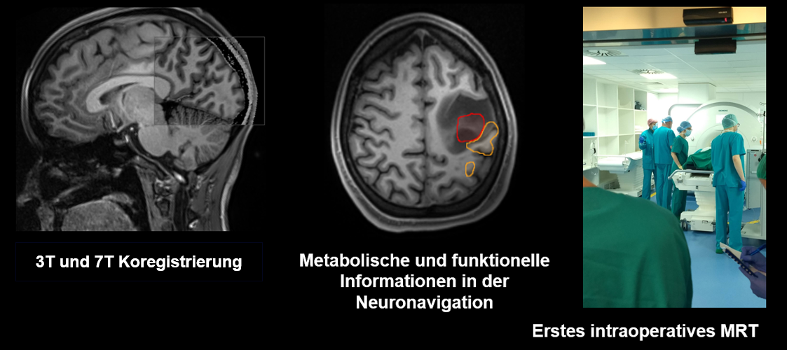
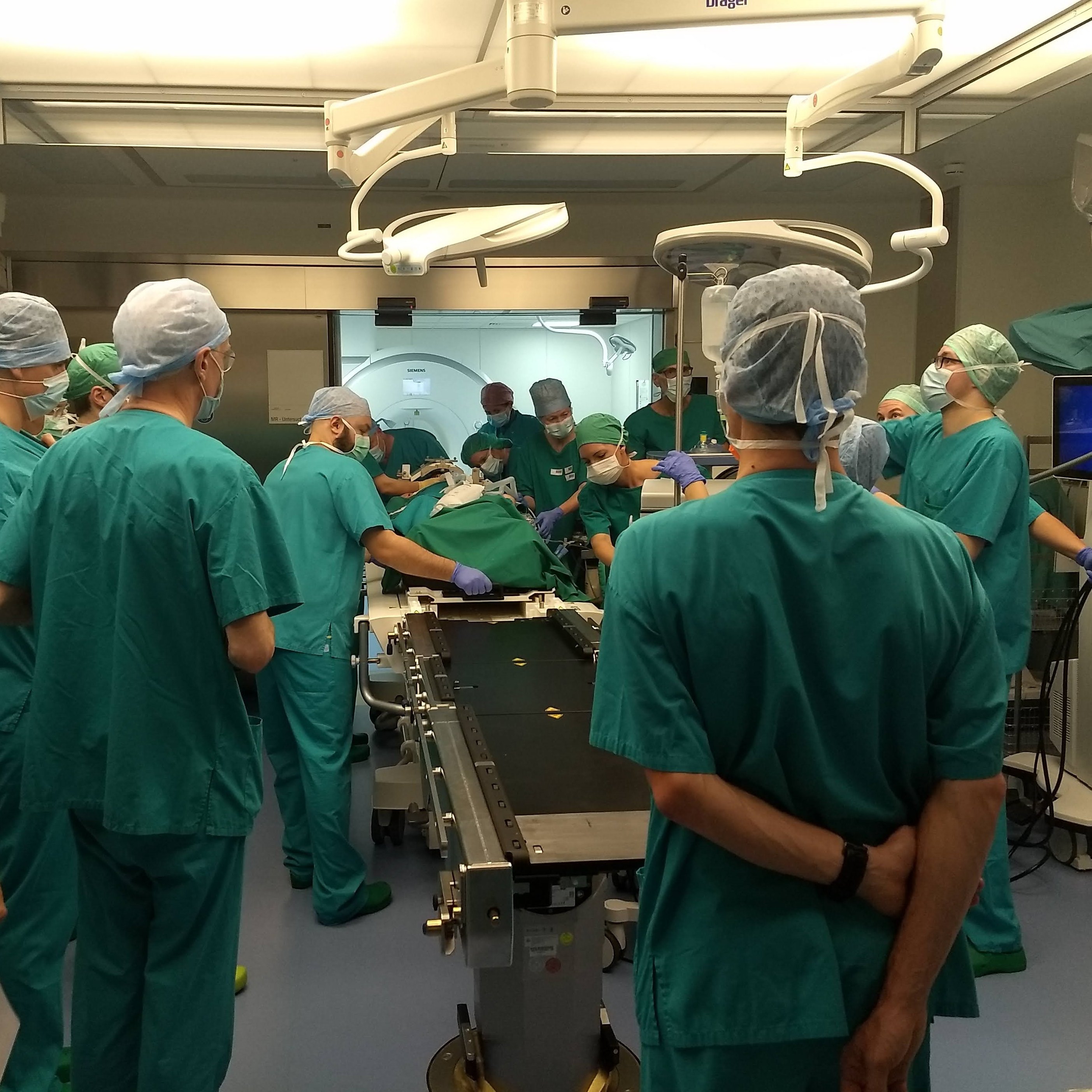
In order to improve imaging not only before but also during surgery, we have an MR device with intraoperative usability, the first in Austria with a field strength of 3 Tesla. This ensures high image quality. Together with modern neuronavigation systems, all available clinical images of the brain can be made available in the operating theater.
Molecular imaging: Ultra-strong magnetic fields for research into brain tumor biology
For many years, the Department of Neurosurgery and the Department of Biomedical Imaging and Image-guided Therapy have been cooperating both in the context of treatment planning for patients and in research projects. One milestone is the joint research into pathologies on the High Field MR Centre’s 7 Tesla scanner.
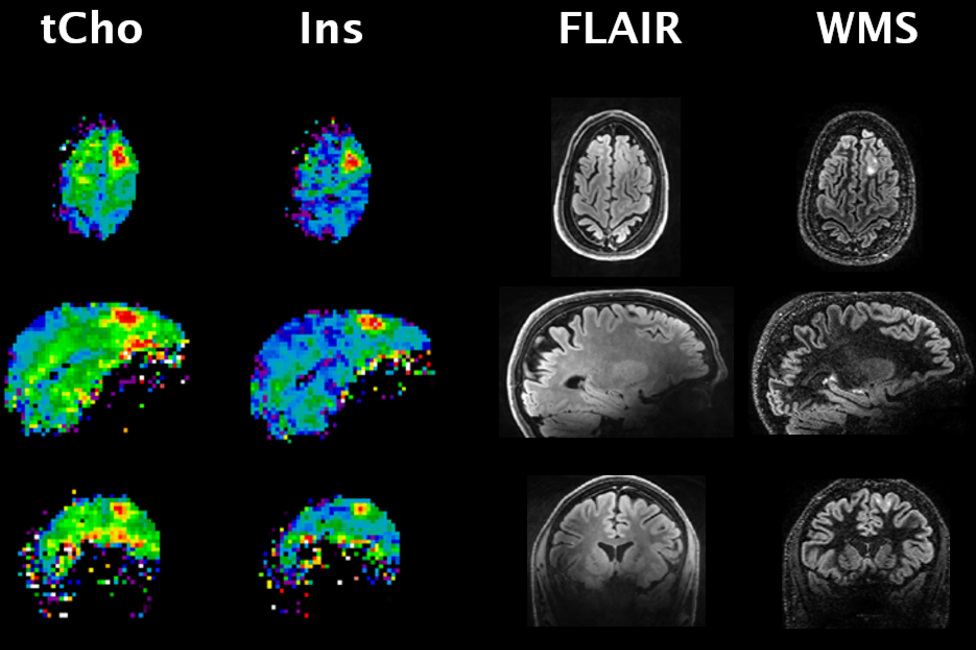
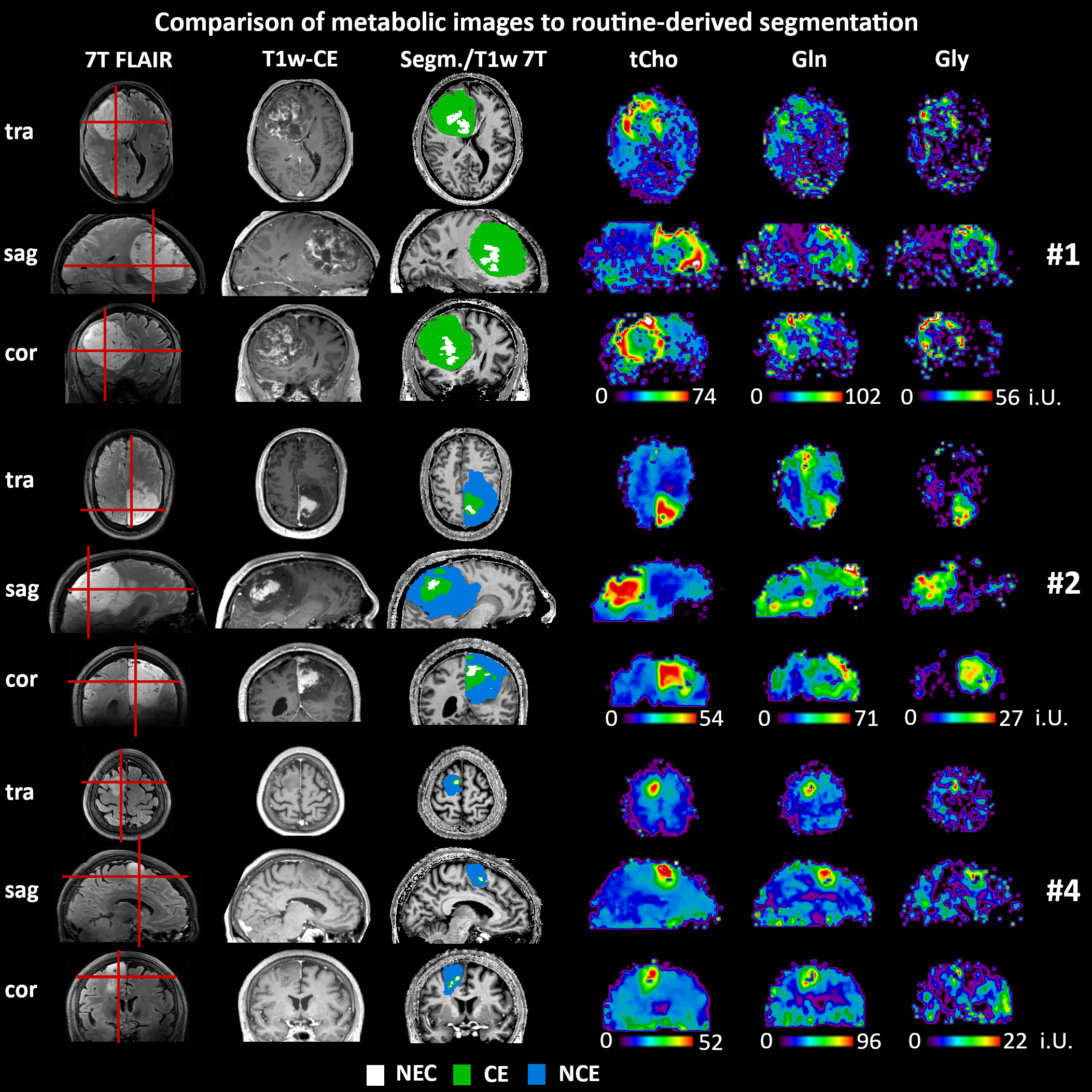
A new method of 7T spectroscopic imaging developed here in Vienna allows the differentiation of metabolites such as glycine and glutamine with high resolutions, which is not possible at low field strengths.
Compared to neuroradiological tumour segmentation, we see strong correlation and different metabolic profiles between necrosis, contrast enhancement and oedema.
A detailed description of our research status can be found in the following brochure: "Meilensteine und Anwendungen der 7 Tesla Magnetresonanz-tomographischen spektroskopischen Bildgebung in Wien" (TEXT AVAILABLE ONLY IN GERMAN!)
Since 2014, latest metabolic imaging based on high-resolution MRSI (magnetic resonance spectroscopy imaging) has been tested in gliomas in collaboration with Prof. Georg Widhalm.
In the past, this research facility was scientifically and practically substantiated as part of an FWF-funded project, "3D 2HG mapping as biomarker for IDH-mutation in glioma".
Since then, gliomas, lymphomas and meningiomas have been measured isotropically using a world-leading 3D MRSI sequence with a resolution of 3.4 mm. First results in high-grade gliomas were successfully published in "NeuroImage: Clinical" in September 2020: „High-resolution metabolic imaging of high-grade gliomas using 7T-CRT-FID-MRSI“.
Since then, we have been working on improved methods and clinically relevant derivations of this technology, for example as part of the FWF project "Quantitative metabolic 7T imaging of tumor microenvironments", which started in 2023. The goals of the project are a better quantitative representation of metabolites, sampling during surgery based on these data and confirmation of metabolic imaging by analytical chemistry and molecular pathology. Furthermore, we will evaluate the potential advantages for therapy planning and monitoring and investigate the correspondence to the current state of the art, such as 5ALA fluorescence.
In 2020, we started studies on epilepsy.
A relevant percentage of people with focal epilepsy cannot be successfully treated with medication and therefore require surgical intervention. The origin of seizures in the brain is not always clear, which is why we want to investigate whether additional information from spectroscopic 7T imaging can lead to better treatment. The preliminary results appear to be promising in terms of elucidating metabolic changes in epilepsy-associated diseases better than ever before.
Prof. Karl Rössler and Gilbert Hangel presenting these studies: “7T MRSI for Epilepsy: Developing a new neurochemical imaging paradigm”
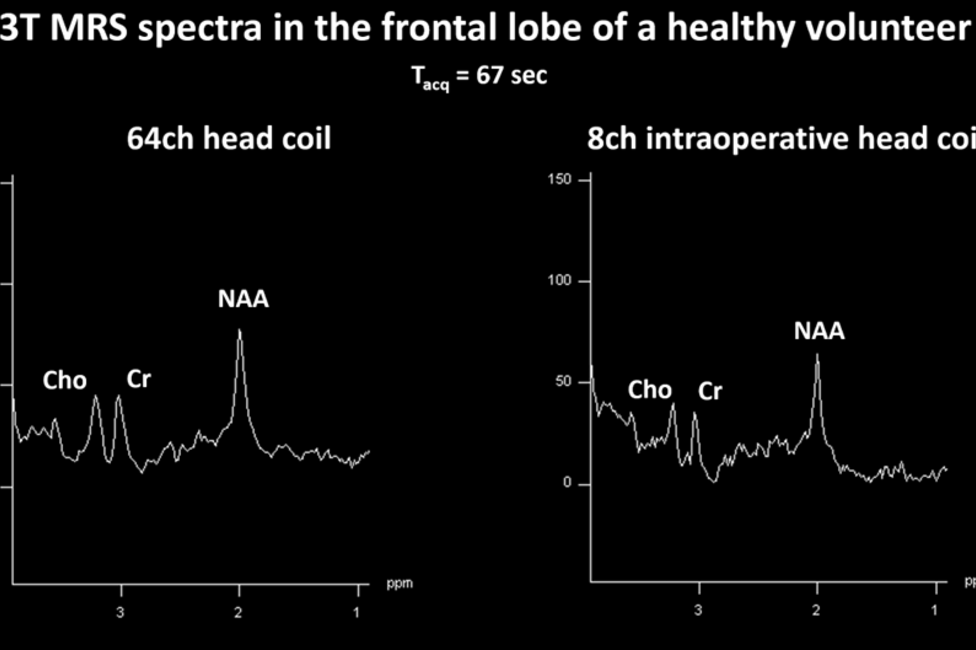
Although intraoperative MR systems are widely used clinically, only structural MRI methods are used as standard in surgery. Based on the expert knowledge of our clinics, we want to develop further advanced methods for visualizing function and metabolism.
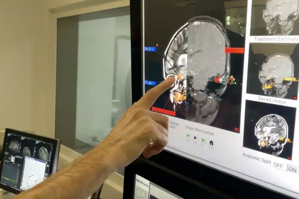
We want to use MR spectroscopy to investigate how tissue behaves after minimally invasive laser-based heating.
Functional imaging can also be performed intraoperatively to image the motor cortex and resting networks in the brain directly before and after a surgical procedure.
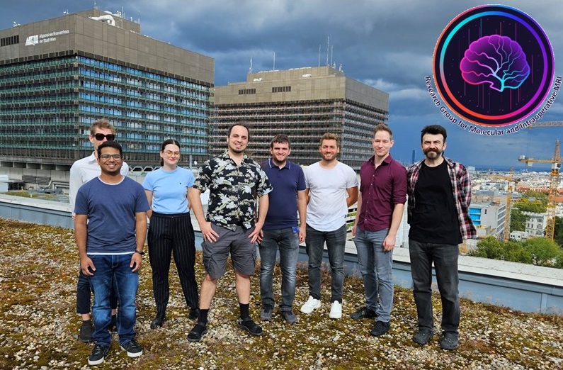
Team of the Molecular and Intraoperative Imaging Working Group
Head:
Priv.-Doz. Dipl.-Ing. Gilbert Hangel, PhD
E-Mail: gilbert.hangel@meduniwien.ac.at
Clinical supervisor:
Univ.-Prof. Dr.med.univ. Karl Rössler
E-Mail: karl.roessler@meduniwien.ac.at
Scientific Staff:
Sagar Acharya - PhD Student
Ahmet Azgin - PhD Student
Cornelius Cadrien - PhD Student
Stefanie Chambers - Studentische Assistentin
Ralph Flandorfer - Studentischer Assistent
Sara Huskic - PhD Studentin
Philipp Lazen - PhD Student
Lorenz Pfleger - PostDoc
Nicolas Weilguny- Studentischer Assistent
Clinical Staff:
Dr.in med.univ.et. scient.med. Barbara Kiesel
Dr.med.univ. et scient.med. Mario Mischkulnig
Dr.in med.univ. Julia Shawarba
Dr. med.univ. Matthias Tomschick
Dr. med.univ. Fabian Winter
Dr. med.univ. Jonathan Wais
Dr. med.univ. Vitalij Zeiser

