Teaching and Training Neurosurgical Operating Techniques
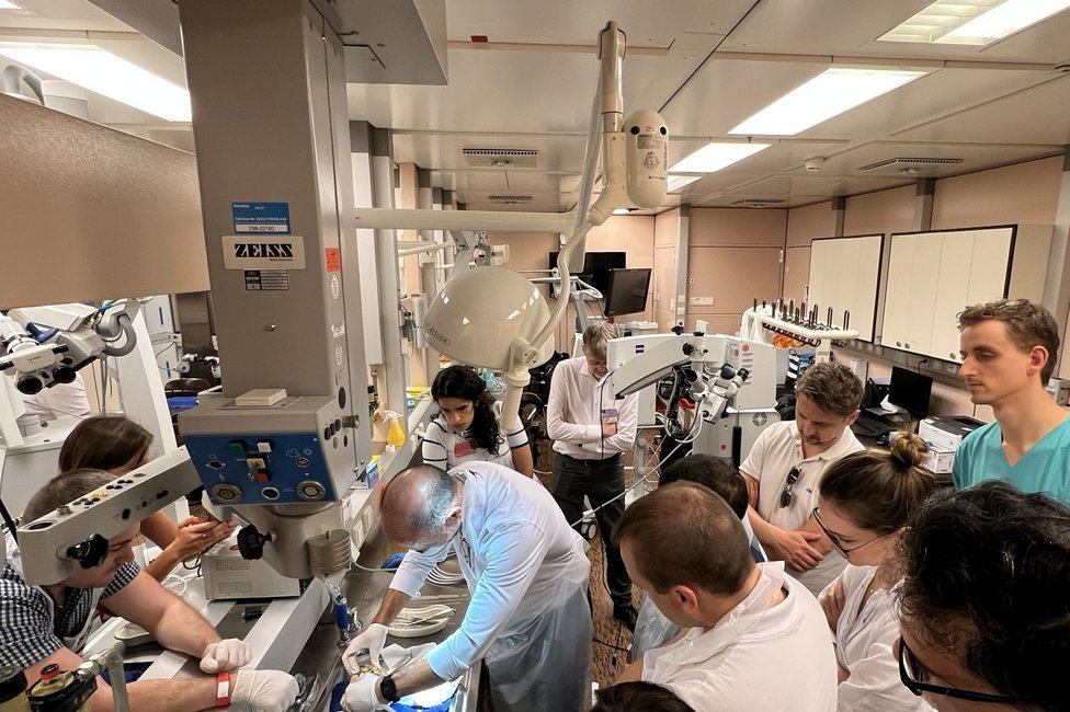
The Microneurosurgery Laboratory on level 8H (AKH Main Building) is used for training and continuing education in neurosurgical operating techniques. Eight workstations, each equipped with a microscope, are routinely available for this purpose. For workshops, the facility can be expanded to up to 16 workstations.
One master workstation is equipped with a high-quality surgical microscope or endoscope with images shared on monitors in the laboratory. A complete set of microneurosurgical instruments is available for training neurosurgical techniques and skills.
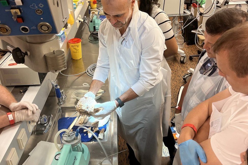
Besides the 40 courses held at regular intervals for participants from a wide range of disciplines (Neurosurgery, Trauma Surgery, Plastic Surgery), microsurgical dissection exercises are offered for students in the form of lectures.
Microneurosurgery Laboratory 8H was also used for demonstration purposes during the citywide event "Long Night of Research".
Innovations in 3D visualisation
A robotic camera system was developed in cooperation with the Austrian Center for Medical Innovation and Technology (ACMIT) for high-resolution, three-dimensional recording of anatomical preparations. As part of the project, the existing surgical microscope was also upgraded with a high-resolution 3D photo and video system. In combination with the 3D projectors at the Department of Neurosurgery, the new set-up now provides a complete 3D system for teaching.
International Workshops and Trainings in the Microneurosurgery Laboratory
Founded 30 years ago by Professor Wolfgang Koos, the "European Workshop on Basic Techniques of Microsurgery and Cerebral Revascularisation" is one of the longest-running microsurgical bypass courses in Europe. The international course, which has already been attended by almost 500 colleagues, is designed to teach different types of microsurgical anastomoses and nerve coaptations. Our programme includes hands-on training on synthetic and animal models, case presentations and lectures by international experts.
To safely perform the Endoscopic Transnasal Surgical Technique, training in anatomic workshops is critical. Hands-on workshops have been held every year since 2003 to provide practical training in endoscopic techniques, or access anatomy with a focus on the endoscopic view of structures.
Twelve fully equipped endoscopy workstations and a 3D endoscopy master station are available in the microsurgical laboratory on level 8H for this course.
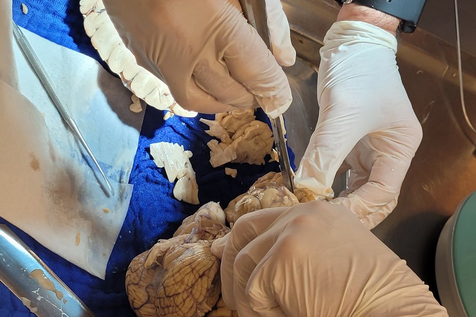
The DTI Workshop has been held 12 times since 2010 to teach participants about the fibre tract anatomy of the brain and deepen their understanding of the correlation with magnetic resonance DTI tractography. Here, the main focus is on the combination of dissection of fibre connections on the cadaver specimen and a virtual dissection of the MRI images using a DTI tractography software. Participants are given the opportunity to familiarise themselves with the anatomical basis of White-Matter-Anatomy and the physical principles and clinical relevance of fibre tract imaging using tractography.


